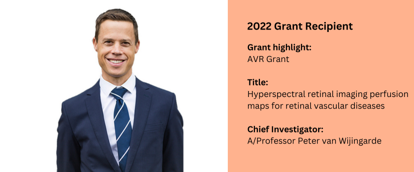
AVR Funded Grant – 2022
Project Title:
Hyperspectral retinal imaging perfusion maps for retinal vascular diseases
Chief Investigator:
A/Professor Peter van Wijingarde
Co-Investigators:
Dr Xavier Hadoux, A/Prof Lyndell Lim, Dr Amy Cohn, Darvy Dang
Aim
The aim of this study was to determine whether hyperspectral imaging could be used to identify areas of retinal ischaemia, as an alternative to fundus fluorescein angiography (FFA) or optical coherence tomography angiography (OCTA).
Methods
Participants: 446 participants were included in the study. 81 participants had retinal ischaemia, identified with either FFA or OCTA imaging. 365 control participants, comprised of a range of healthy people, those with glaucoma or intermediate age- related macular degeneration with no evidence of retinal ischemia, were also included in the study. All study participants underwent retinal hyperspectral imaging. The foveal avascular zone of control participants was utilised as an internal control for avascular parameter comparison.
Key results
Overall Agreement:
• The model demonstrated high performance for the detection of retinal ischemia using hyperspectral images alone.
Ischemia Classification:
• Of a total 81 participants with known ischemia, the model correctly identified ischemia in 74 cases (91.4%).
Non ischemic Control Classification:
• In the control group (N=365), the model identified non-perfusion limited to the foveal area in 321 cases (87.9%)
Implications for Clinical Practice/Science and Future Research
We are in the process of developing an independent data set to validate and, if necessary, further refine our deep learning model. It is our expectation that the results of this study and the subsequent validation will be published and the subject of scientific presentations.
Conclusion
We have successfully developed a deep learning model for the analysis of retinal hyperspectral images for the detection of retinal ischaemia. The model was developed to process spectral (multiple wavelength) and spatial information. The model is sensitive and specific (robust to comorbid retinal pathology) and may have promise in the detection of retinal ischaemia in primary eye care settings, or in specialist care as a precursor to OCTA or FFA.
Lay Summary of Outcomes
We have shown that a new type of retinal photography with a rainbow-coloured flash can be used to detect areas of the retina that are not getting enough blood supply. Detecting impaired retinal blood supply can enable timely treatment of diabetic retinopathy and save sight.

